Current
location:
Current
location: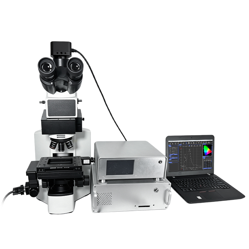

Product Introduction
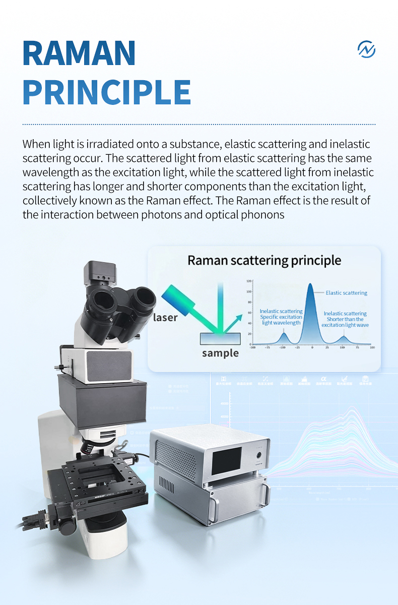
Pharmaceutical quality control, biomedical research, and forensic science share a common analytical challenge: the samples they study are overwhelmingly organic, colored, or biologically derived — precisely the materials that generate intense autofluorescence under visible laser excitation. A 532 nm laser striking a pharmaceutical tablet, tissue section, or dyed textile fiber typically produces a broad fluorescence background that swamps the much weaker Raman signal, rendering spectral interpretation difficult or impossible. This fluorescence problem is not a minor inconvenience; it is the single largest barrier to adopting Raman spectroscopy in pharmaceutical, biomedical, and forensic laboratories.
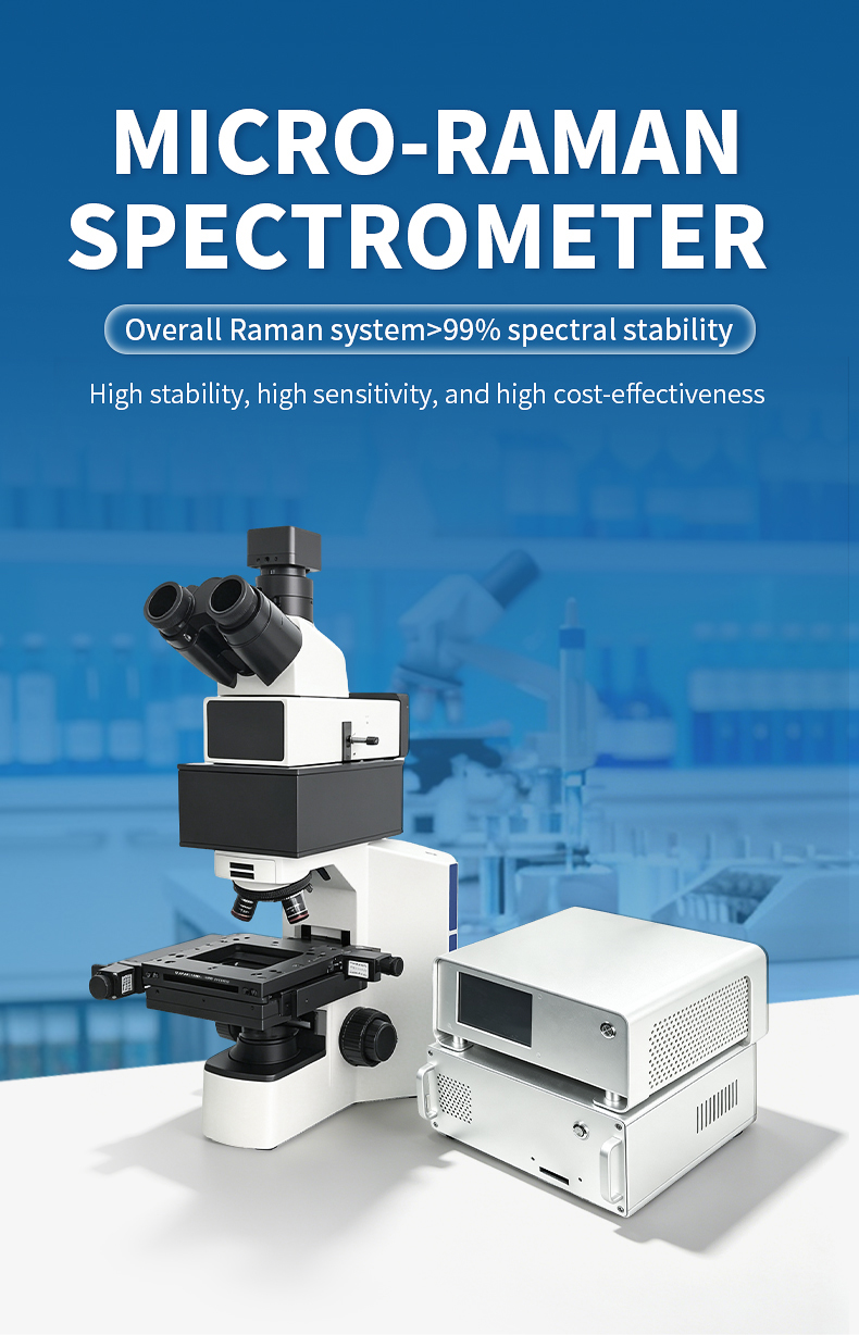
The MR-785A micro Raman spectrometer was designed specifically to overcome this barrier. By employing a 785 nm near-infrared excitation laser, the system operates at a wavelength where most organic and biological materials exhibit minimal autofluorescence. The physics is straightforward: fluorescence requires absorption of the excitation photon, and most organic chromophores do not absorb at 785 nm. The result is clean, interpretable Raman spectra from samples that would produce featureless fluorescence backgrounds under 532 nm excitation.
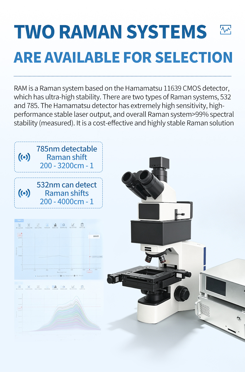
The optical system pairs a 1200 gl/mm volume phase holographic grating blazed at 800 nm with the Hamamatsu 11639 CMOS detector (2048 pixels, >99.8% linearity), delivering a wavenumber range of 200–3200 cm⁻¹ with 7 cm⁻¹ spectral resolution. This configuration captures the diagnostically critical fingerprint region where pharmaceutical polymorphs exhibit their most distinctive spectral differences, where biological tissues show characteristic protein, lipid, and nucleic acid bands, and where forensic fibers reveal dye composition signatures.
For pharmaceutical laboratories performing raw material verification, polymorph screening, or finished product inspection, the MR-785A provides a non-destructive identification tool that works through blister packaging and glass vials. For biomedical researchers studying tissue biochemistry, the system reveals protein secondary structure, lipid composition, and nucleic acid content without staining or sectioning. For forensic scientists, it enables non-destructive fiber comparison and dye identification without removing evidence from its collection substrate. Read our Raman spectrometer value comparison to see how the MR-785A stacks up against alternatives.
Applications
- Pharmaceutical polymorph identification — Distinguish drug crystal forms (polymorphs I, II, III, etc.) that differ in bioavailability and patent status, using fingerprint region spectral comparison without sample destruction
- Raw material identity verification through packaging — Confirm incoming API and excipient identity by measuring through glass vials and transparent bags, eliminating contamination risk from container opening
- Biological tissue biochemistry profiling — Map protein, lipid, and nucleic acid distributions in unstained tissue sections for cancer research, pathology, and developmental biology studies
- Forensic fiber comparison and dye identification — Non-destructively compare textile fibers by polymer type and dye composition, supporting trace evidence analysis in criminal investigations
- Drug substance and counterfeit detection — Identify unknown tablets, capsules, and powders by spectral library matching, supporting law enforcement and pharmaceutical authentication
- Food safety contaminant screening — Detect melamine, Sudan dyes, pesticide residues, and other organic contaminants on food surfaces with characteristic Raman fingerprint identification
- Geological fluid inclusion analysis — Characterize trapped fluids and gases in mineral inclusions where the host mineral fluoresces under visible excitation
- Art conservation and heritage science — Identify organic pigments, binders, varnishes, and degradation products on historical artifacts where fluorescence is inherent to the aged materials
Key Features and Advantages
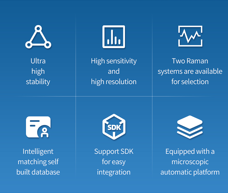
- 785 nm NIR excitation suppresses autofluorescence from organic compounds, biological tissues, dyed fibers, and colored minerals by orders of magnitude compared to visible-wavelength lasers
- 1200 gl/mm grating blazed at 800 nm maximizes diffraction efficiency at the excitation wavelength, compensating for the inherently weaker Raman scattering at longer wavelengths
- Hamamatsu CMOS detector with 2048 pixels and >99.8% linearity enables quantitative peak intensity comparison essential for polymorph ratio analysis and tissue biochemistry quantification
- 200–3200 cm⁻¹ wavenumber range covers the complete fingerprint region where pharmaceutical polymorphs, biological molecules, and forensic dyes exhibit their most distinctive spectral features
- 7 cm⁻¹ spectral resolution resolves polymorph-specific peak positions and bandwidths, enabling discrimination between closely related crystal forms (e.g., ritonavir Forms I and II)
- Signal-to-noise ratio of 500:1 ensures interpretable spectra from weak organic Raman scatterers that produce inherently lower signal than crystalline inorganic materials
- Non-contact measurement through transparent packaging (glass, clear plastic, blister packs) enables sealed-container identification for pharmaceutical receiving and hazardous material screening
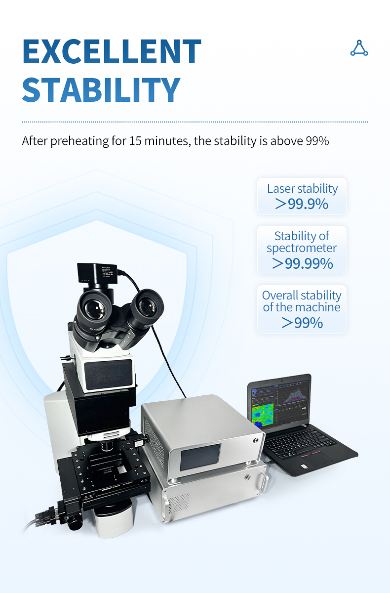
- System stability >99% over 8 hours supports batch analysis of dozens of pharmaceutical samples or forensic evidence items without recalibration interruption
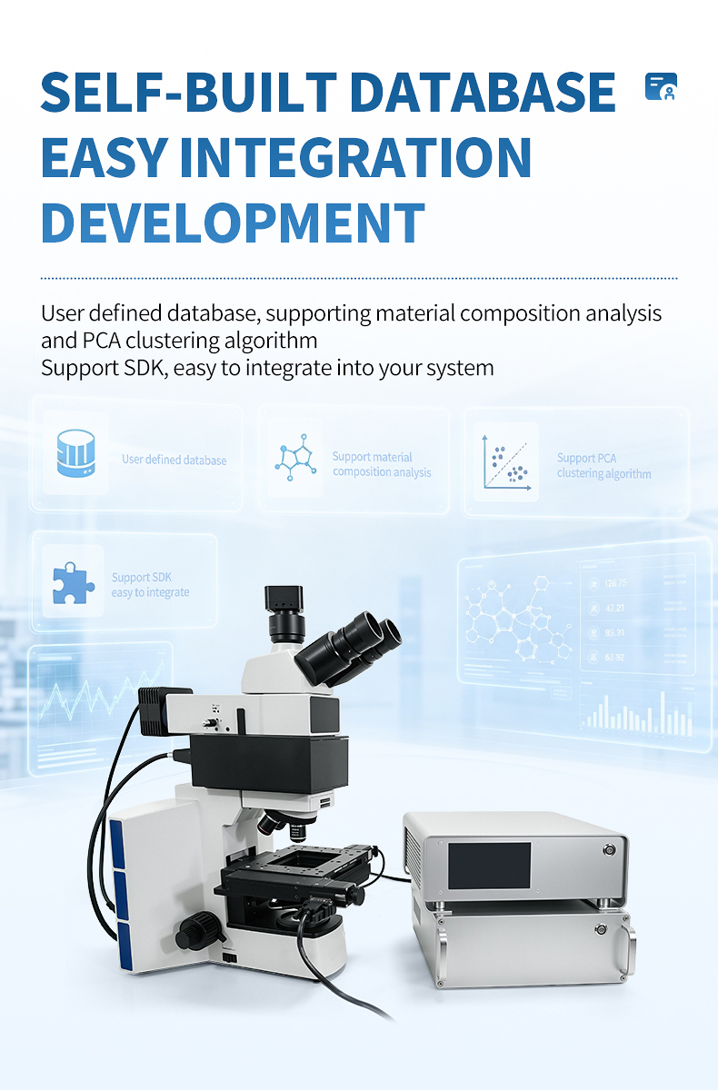
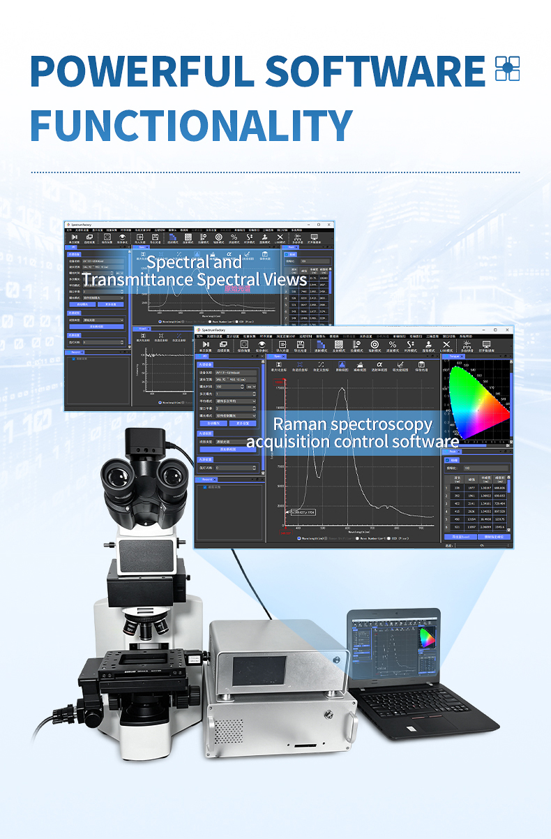
- SpectrumFactory software with built-in pharmaceutical and forensic spectral libraries enables rapid compound identification with match quality scoring
- PCA clustering analysis automatically groups similar spectra within batch datasets, supporting polymorph screening and tissue region classification without manual spectral interpretation
- airPLS baseline correction handles residual fluorescence background that may appear even at 785 nm, ensuring clean spectra for reliable database matching
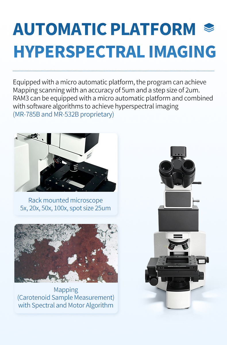
- Rack-mounted microscope with 5x–100x objectives and 25 μm spot size enables targeted analysis of individual pharmaceutical particles, single fibers, and micro-tissue regions
- SMA905 fiber optic interface supports future system upgrades and integration with external sampling accessories for specialized applications
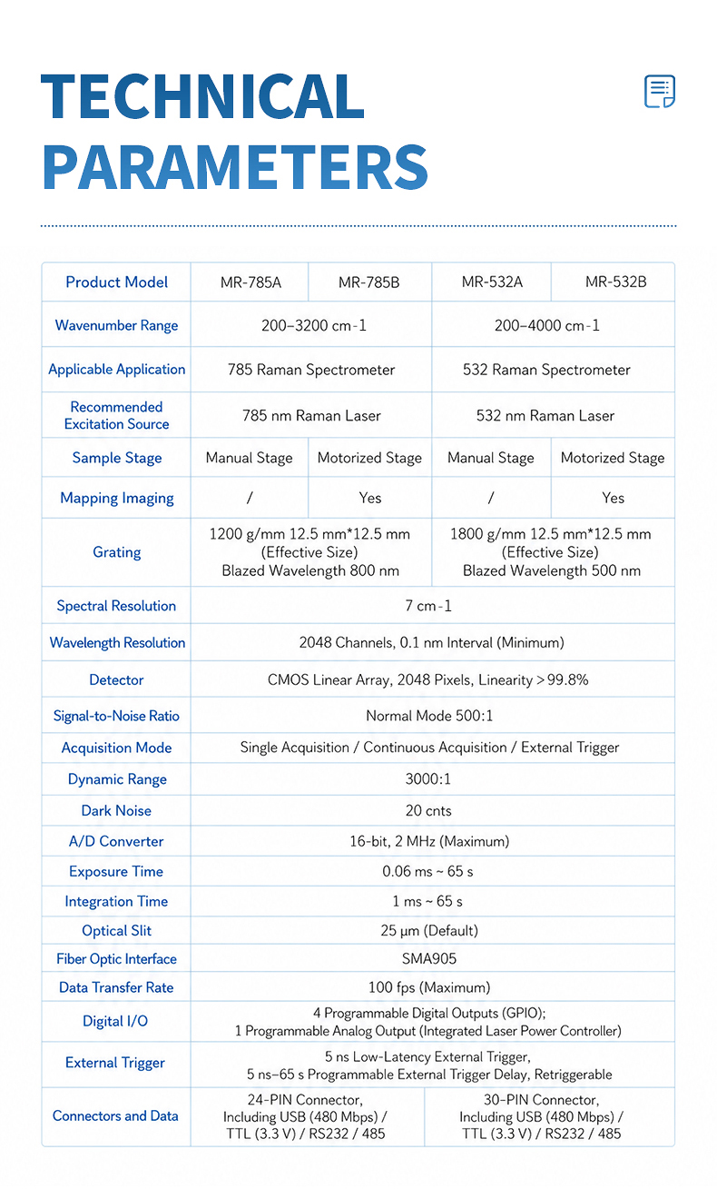
Technical Specifications
| Parameter | Specification |
|---|---|
| Product Model | MR-785A |
| Excitation Wavelength | 785 nm |
| Wavenumber Range | 200–3200 cm⁻¹ |
| Spectral Resolution | 7 cm⁻¹ |
| Grating | 1200 gl/mm, 12.5 mm × 12.5 mm, blazed at 800 nm |
| Detector | Hamamatsu CMOS linear array, 2048 pixels, linearity > 99.8% |
| Wavelength Resolution | 2048 channels, 0.1 nm interval (minimum) |
| Signal-to-Noise Ratio | 500:1 (normal mode) |
| Dynamic Range | 3000:1 |
| Dark Noise | 20 counts |
| A/D Converter | 16-bit, 2 MHz (maximum) |
| Acquisition Mode | Single / Continuous / External trigger |
| Exposure Time | 0.06 ms – 65 s |
| Integration Time | 1 ms – 65 s |
| Optical Slit | 25 μm (default) |
| Sample Stage | Manual platform |
| Microscope | Rack-mounted, 5x / 20x / 50x / 100x objectives |
| Spot Size | 25 μm |
| Fiber Optic Interface | SMA905 |
| Data Transfer Rate | 100 fps (maximum) |
| Digital I/O | 4 programmable GPIO; 1 analog output |
| External Trigger | 5 ns latency, 5 ns–65 s delay, retriggerable |
| Connectors | 30-pin (USB 480 Mbps / TTL 3.3 V / RS232 / RS485) |
| System Stability | Laser >99.9%, Spectrometer >99.99%, Overall >99% (8h after 15-min warm-up) |
FAQ
Q: Why is 785 nm better than 532 nm for pharmaceutical Raman analysis?
A: Most pharmaceutical compounds contain organic chromophores that absorb visible light, producing fluorescence that overwhelms Raman signals at 532 nm. At 785 nm, photon energy is below most chromophores' absorption threshold, virtually eliminating fluorescence. Although signal is approximately 3.5x weaker, the absence of fluorescence yields much higher signal-to-background ratios. 785 nm is the industry standard for pharmaceutical Raman identification.
Q: Can the MR-785A distinguish between drug polymorphs?
A: Yes. Different polymorphs exhibit distinct Raman spectra due to differences in crystal packing and molecular conformation. The 7 cm⁻¹ resolution resolves polymorph-specific peak positions and bandwidths. The database matching function stores reference spectra and performs automated identification with quality scoring.
Q: How effective is the MR-785A for analyzing biological tissue samples?
A: The 785 nm excitation is ideal for label-free tissue analysis. Tissues show characteristic Raman bands from proteins (amide I/III), lipids (CH₂/C=C), and nucleic acids. Low autofluorescence at 785 nm reveals these signatures clearly, enabling tissue classification and tumor margin assessment.
Q: Can I use the MR-785A for forensic fiber analysis without destroying evidence?
A: Absolutely. Raman is inherently non-destructive and non-contact. The MR-785A measures fibers directly on evidence tape or in bags without sample removal. 785 nm minimizes dye fluorescence, enabling identification of both fiber polymer type and dye composition. The fiber remains intact for DNA analysis or courtroom presentation.
Q: What integration times are typically needed for organic and pharmaceutical samples at 785 nm?
A: Organic and pharmaceutical samples typically need 2–10 seconds for concentrated tablets, 5–30 seconds for biological tissues, and 10–60 seconds for dilute samples. The 1 ms to 65 s integration range covers all scenarios, with airPLS handling residual fluorescence.
Q: Does the MR-785A comply with pharmaceutical industry regulatory requirements?
A: The hardware and SpectrumFactory software support pharmaceutical QC workflows. The system calibrates against pharmacopeial standards, and through-package measurement supports cGMP receiving protocols. Software audit trails and access controls can be configured for 21 CFR Part 11 compliance.
Article address:https://www.spectrometer.top/micro-raman-spectrometer/raman-pharmaceutical-biomedical-forensic.html